Finecare RF (Rheumatoid Factor) Rapid Quantitative Test kit
Finecare SAA (Serum Amyloid A) Rapid Quantitative Test -immunoassay
Key Features and Specifications
-
Test Principle: Fluorescence immunoassay.
-
Sample Type: Serum, plasma, or whole blood.
-
Measuring Range: Typically 3 – 300 mg/L (varies by analyzer model).
-
Reaction Time: Approximately 15 minutes.
-
Sample Volume: Around 75 µL.
-
Storage Temperature: 4°C – 30°C.
-
Shelf Life: Valid until the expiration date indicated on the packaging.
-
Compatible Device: Finecare FIA Analyzer (e.g., Finecare FS-113, Finecare Plus).
Finecare T3 (Triiodothyronine) Control Kit
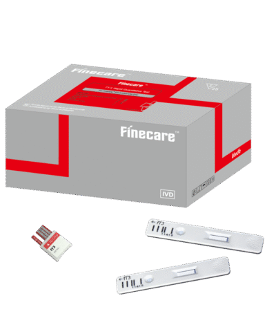
Finecare T3 Rapid Quantitative Test
✅ Key Features & Specifications
-
Test principle: Fluorescence Immunoassay (FIA) for the quantitative measurement of Total Triiodothyronine (T3) in human serum, plasma, or whole blood.
-
Analyte: Total T3 (TT3).
-
Measurement range: Typically 0.3–8.0 ng/mL (may vary slightly depending on the lot).
-
Sample types: Whole blood, serum, or plasma.
-
Sample volume: Approximately 75–100 µL per test.
-
Turnaround time: Results available in 15 minutes.
-
Storage conditions: Test cartridges stable at 4 °C – 30 °C, with a shelf life up to 24 months.
-
Instrument compatibility: Requires a Finecare™ Fluorescence Immunoassay Analyzer (Finecare, Finecare Plus, or Finecare II) for quantitative reading.

Finecare T4 Rapid Quantitative Test immunoassay
✅ Key Features & Specifications
-
Test principle: Fluorescence Immunoassay (FIA) for the quantitative measurement of Total Thyroxine (T4) in human serum, plasma, or whole blood.
-
Analyte: Total T4 (TT4).
-
Measurement range: Typically 2–18 µg/dL (may vary slightly depending on lot).
-
Sample types: Whole blood, serum, or plasma.
-
Sample volume: Approximately 75–100 µL per test.
-
Turnaround time: Results available in 15 minutes.
-
Storage conditions: Test cartridges stable at 4 °C – 30 °C, with a shelf life up to 24 months.
-
Instrument compatibility: Requires a Finecare™ Fluorescence Immunoassay Analyzer (Finecare, Finecare Plus, or Finecare II) for quantitative reading.
Finecare Testosterone Rapid Quantitative Test Kit immunoassay
Key Features and Specifications
-
Test Principle: Fluorescence immunoassay.
-
Sample Type: Serum, plasma, or whole blood.
-
Measuring Range: Typically 0.1 – 15 ng/mL (may vary with analyzer model).
-
Reaction Time: Approximately 15 minutes.
-
Sample Volume: About 75 µL.
-
Storage Temperature: 4°C – 30°C.
-
Shelf Life: Valid until the indicated expiry date.
-
Compatible Analyzer: Finecare FIA Analyzer (e.g., Finecare FS-113, Finecare Plus).
Finecare Total IgE Rapid Quantitative Test Kit immunoassay
Key Features and Specifications
-
Test Principle: Fluorescence immunoassay.
-
Sample Type: Serum, plasma, or whole blood.
-
Measuring Range: Typically 10 – 2000 IU/mL (depending on analyzer model).
-
Reaction Time: Approximately 15 minutes.
-
Sample Volume: About 75 µL.
-
Storage Temperature: 4°C – 30°C.
-
Shelf Life: Up to the indicated expiry date.
-
Compatible Device: Finecare FIA Analyzer (e.g., Finecare FS-113, Finecare Plus).

Finecare TSH Rapid Quantitative Test immunoassay
✅ Key Features & Specifications
-
Test principle: Fluorescence Immunoassay (FIA) for the quantitative measurement of Thyroid Stimulating Hormone (TSH) in human serum, plasma, or whole blood.
-
Analyte: Thyroid Stimulating Hormone (TSH).
-
Measurement range: Typically 0.1–100 µIU/mL (may vary slightly depending on the lot).
-
Sample types: Whole blood, serum, or plasma.
-
Sample volume: Approximately 75–100 µL per test.
-
Turnaround time: Results available in 15 minutes.
-
Storage conditions: Test cartridges stable at 4 °C – 30 °C, with a shelf life up to 24 months.
-
Instrument compatibility: Requires a Finecare™ Fluorescence Immunoassay Analyzer (Finecare, Finecare Plus, or Finecare II) for quantitative reading.
Finecare TSH/T3/T4 Control Kit
Finecare Vitamin B12 Rapid Quantitative Test Kit
Finecare Vitamin D Rapid Quantitative immunoassay
✅ Key Features & Specifications
-
Test principle: Fluorescence Immunoassay (FIA) for the quantitative measurement of 25-hydroxy Vitamin D [25(OH)D] in human serum, plasma, or whole blood.
-
Analyte: 25-hydroxy Vitamin D (25(OH)D).
-
Measurement range: Typically 4–100 ng/mL (may vary slightly depending on the lot).
-
Sample types: Whole blood, serum, or plasma.
-
Sample volume: Approximately 75–100 µL per test.
-
Turnaround time: Results available in 15 minutes.
-
Storage conditions: Test cartridges stable at 4 °C – 30 °C, with a shelf life up to 24 months.
-
Instrument compatibility: Requires a Finecare™ Fluorescence Immunoassay Analyzer (Finecare, Finecare Plus, or Finecare II) for quantitative reading.