
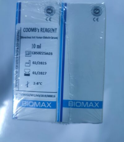
BIOMAX Coomb’s Reagent
Original price was: KSh 2,000.00.KSh 1,500.00Current price is: KSh 1,500.00.A laboratory reagent used in blood testing to detect antibodies bound to red blood cells. It is essential for performing the Coombs test (direct and indirect), which helps identify blood compatibility issues and immune-related blood disorders. Reliable and easy to use, suitable for hospitals, clinics, and blood banks.
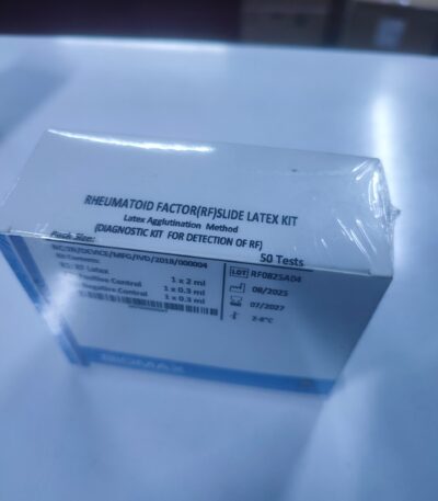
Biomax Rheumatoid Factor (RF) Slide Latex Kit
Original price was: KSh 1,800.00.KSh 1,200.00Current price is: KSh 1,200.00. Order via WhatsApp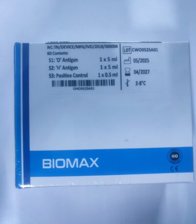
Biomax Widal Antigen Test Kit
Original price was: KSh 2,000.00.KSh 1,100.00Current price is: KSh 1,100.00. Order via WhatsApp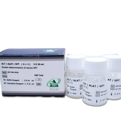
Biotech: ALAT / ALT / GPT Reagents — Clinical Chemistry / Semi-Automated Systems-biochemistry
Original price was: KSh 5,000.00.KSh 3,600.00Current price is: KSh 3,600.00.🔬 1. IFCC Kinetic Method (Standard ALT Method)
Primary Reaction
ALT catalyzes:
L-Alanine + α-Ketoglutarate
→ Pyruvate + L-Glutamate
Indicator (Coupled) Reaction
Pyruvate + NADH + H⁺
→ Lactate + NAD⁺
(catalyzed by LDH)
As ALT activity increases, NADH decreases, and absorbance at 340 nm falls.
Measurement
-
Wavelength: 340 nm
-
Method: Kinetic, ΔA/min
-
Rate proportional to ALT activity (U/L)
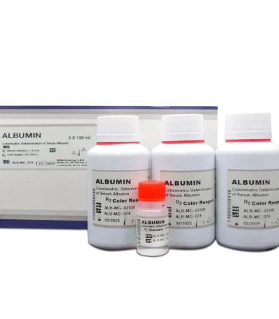
Biotech: Albumin Reagents- biochemistry
Original price was: KSh 5,000.00.KSh 3,500.00Current price is: KSh 3,500.00.🔬 1. BCG Method (Bromocresol Green)
Most widely used in routine chemistry analyzers.
Principle
Albumin binds to BCG dye in an acidic buffer forming a green-colored complex.
-
Absorbance: 600–630 nm (green complex)
-
Type: Endpoint reaction (color intensity ∝ albumin concentration)
Advantages
-
Very robust
-
Strong signal (high sensitivity)
Limitations
-
Mild positive bias in some conditions (e.g., high globulins)
🧪 2. BCP Method (Bromocresol Purple)
A more specific albumin method.
Principle
Albumin binds selectively to BCP dye in acidic buffer forming a purple complex.
-
Absorbance: 540–550 nm
-
Type: Endpoint
Advantages
-
Higher specificity than BCG
-
Less interference from globulins or acute phase proteins
Limitations
-
Slightly lower color intensity
-
More common in premium reagent lines
Biotech: Alkaline Phosphatase (ALP / ALKP) Reagents biochemistry
Original price was: KSh 5,000.00.KSh 3,600.00Current price is: KSh 3,600.00.🔬 1. IFCC Kinetic ALP Method (Standard)
Principle
ALP + p-Nitrophenyl phosphate (pNPP)
→ p-Nitrophenol (yellow) + Phosphate
-
Absorbance increases as p-nitrophenol is formed.
-
The rate of increase (ΔA/min) at 405 nm is proportional to ALP activity.
Measurement
-
Wavelength: 405 nm
-
Mode: Kinetic (multiple interval readings)
-
Temperature: 37°C (IFCC standard)
🧪 2. Reagent Composition (Typical)
Most ALP kits use single-reagent or two-reagent formats.
R1 (Main Buffer Reagent)
-
Diethanolamine (DEA) buffer (high pH ~10.3)
-
Magnesium ions (Mg²⁺) — ALP cofactor
-
Zinc ions (Zn²⁺) — enzyme stabilizer
-
Surfactants
-
Preservatives
R2 (Substrate Reagent)
-
p-Nitrophenyl phosphate (pNPP)
-
Stabilizers
-
Preservatives
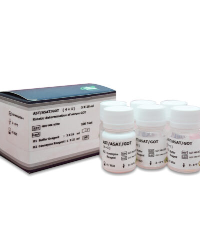
Biotech: ASAT / AST / SGOT Reagents — Clinical Chemistry / Semi-Automated Systems-biochemistry
Original price was: KSh 5,000.00.KSh 3,600.00Current price is: KSh 3,600.00.🔬 1. IFCC Kinetic Method (Standard AST Method)
Primary Reaction (AST enzyme reaction)
L-Aspartate + α-Ketoglutarate
→ Oxaloacetate + L-Glutamate
Indicator (Coupled) Reaction
Oxaloacetate + NADH + H⁺
→ Malate + NAD⁺
(catalyzed by MDH)
LDH is often included to convert pyruvate → lactate, preventing inhibition.
As AST activity increases, NADH decreases, causing a fall in absorbance at 340 nm.
Measurement
-
Wavelength: 340 nm
-
Mode: Kinetic (rate), ΔA/min
-
Rate proportional to AST activity (U/L)
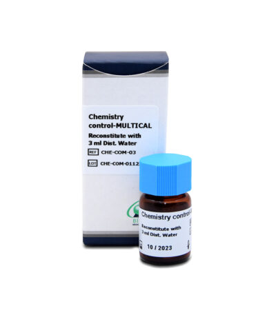
Biotech: Biochemistry Reagents
Original price was: KSh 5,000.00.KSh 3,500.00Current price is: KSh 3,500.00.1. Diagnostic Purpose
Biochemistry reagents are designed to quantitatively measure analytes such as:
-
Liver function markers (ALT, AST, ALP, GGT, Bilirubin, Total Protein)
-
Renal function markers (Urea, Creatinine, Uric Acid)
-
Electrolytes (Sodium, Potassium, Chloride—depending on analyzer type)
-
Lipid profile (Cholesterol, Triglycerides, HDL, LDL)
-
Glucose metabolism (Glucose, HbA1c kits depending on system)
-
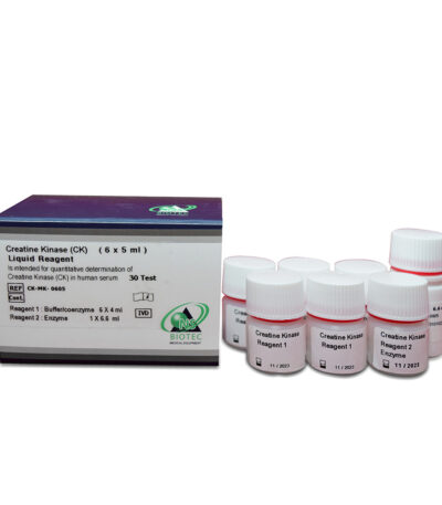
Enzymes and substrates (LDH, CK, Amylase, Lipase)
2. Types of Formulations
Biochemistry reagents may come in:
-
Single-reagent systems
(One ready-to-use bottle) -
Two-reagent systems
(R1 buffer + R2 starter/working reagent) -
Lyophilized reagents
(Require reconstitution) -
Liquid-stable reagents
(Long shelf-life, preferred for automated analyzers)
3. Compatibility
Designed for:
-
Fully automated clinical chemistry analyzers
-
Semi-automated bench-top analyzers
-
Point-of-care analyzers (specific formats)
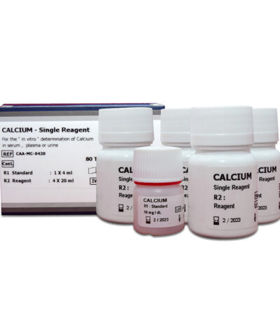
Biotech: Calcium Reagents-biochemistry
Original price was: KSh 5,000.00.KSh 3,300.00Current price is: KSh 3,300.00.Principle of Measurement
1. Arsenazo III Method (Most widely used in modern analyzers)
How it works:
-
Calcium ions react with Arsenazo III dye to form a blue-purple complex.
-
The intensity of the color is measured photometrically at 650–660 nm.
-
Highly specific for calcium with minimal interference from magnesium.
Advantages:
-
Excellent stability
-
Strong absorbance
-
High specificity and precision
2. o-Cresolphthalein Complexone (OCPC) Method
How it works:
-
Calcium forms a purple-red complex with o-cresolphthalein complexone in alkaline conditions.
-
Read photometrically at 570–580 nm.
-
Common in semi-auto chemistry analyzers.
Advantages:
-
Reliable and widely established
-
Suitable for manual and automated systems
Reagent Components
Arsenazo III Reagents May Contain:
-
Arsenazo III dye
-
Buffer (MES or other suitable buffers)
-
Stabilizers
-
Surfactants
-
Preservatives
OCPC Reagents May Contain:
-
o-Cresolphthalein complexone
-
Alkaline buffer (e.g., 8-hydroxyquinoline to remove magnesium interference)
-
Dye stabilizer
-
Surfactants
-
Preservatives
Packaging Format
-
Single liquid reagent (most common)
-
Two-reagent system (R1 + R2)
-
Ready-to-use liquid stable bottles
-
Analyzer-specific reagent cartridges
Common volumes: 25 ml, 50 ml, 100 ml, 250 ml or more.
Sample Types
-
Serum
-
Plasma (heparinized samples preferred)
-
Urine (dilution may be needed)
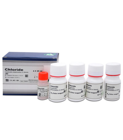
Biotech: Chloride Reagents-biochemistry
Original price was: KSh 5,000.00.KSh 3,600.00Current price is: KSh 3,600.00.Measurement Methods & Reagents
1. Ion-Selective Electrode (ISE) Method (Most common in modern analyzers)
Reagents include:
-
Reference (internal standard) solution
-
Electrode electrolyte solution
-
Conditioning solution
-
Cleaning solution
-
Calibrators (Low and High Chloride)
Principle:
A chloride-selective membrane develops an electrical potential proportional to chloride concentration.
ISE systems require stable calibration solutions and maintenance reagents.
2. Mercuric Thiocyanate Colorimetric Method
How it works:
-
Chloride reacts with mercuric ions, forming mercuric chloride.
-
Released thiocyanate ions form a red-colored complex with ferric ions.
-
The color intensity is measured photometrically at 480–520 nm.
Reagents contain:
-
Mercuric thiocyanate
-
Ferric nitrate or ferric ammonium sulfate
-
Buffer system
-
Surfactants and stabilizers
Advantages:
-
Reliable for automated chemistry analyzers
-
High precision
3. Colorimetric TPTZ Method (Less common)
Chloride displaces iron from a complex; the released iron reacts with TPTZ dye to form a measurable color.
Packaging Formats
-
Single-liquid reagent
-
Two-reagent kits (R1 buffer + R2 color reagent)
-
ISE-specific reagent packs
-
Ready-to-use liquid stable formulations
Typical volumes: 25 ml, 50 ml, 100 ml, 250 ml.
Biotech: Creatinine Reagents
Original price was: KSh 5,000.00.KSh 3,500.00Current price is: KSh 3,500.00.1. Jaffe Method (Most Common)
-
Based on the reaction between creatinine and picric acid in an alkaline medium.
-
Produces an orange-red complex.
-
Absorbance is measured photometrically, usually at 505–520 nm.
-
Some kits include compensated Jaffe formulations to reduce interference from glucose, ketones, and proteins.
2. Enzymatic Method
-
Uses a sequence of enzyme reactions (creatininase, creatinase, sarcosine oxidase, peroxidase).
-
Produces a colored dye measurable at 550–570 nm.
-
Higher specificity than Jaffe, minimal interference.
Reagent Components
Depending on the method, creatinine reagent kits may include:
For Jaffe Method
-
Alkaline buffer
-
Picric acid
-
Stabilizers
-
Surfactants
-
Preservatives
For Enzymatic Method
-
Enzyme mixture (creatininase, creatinase, sarcosine oxidase)
-
Chromogenic dye reagents
-
Phosphate or Good’s buffer
-
Activators
-
Preservatives
Packaging Format
Most creatinine reagent kits are provided as:
-
Two-reagent system (R1 buffer + R2 working reagent)
-
Ready-to-use liquid formulations
-
Optional standards or calibrators
Common sizes: 25 ml, 50 ml, 100 ml, 250 ml, or analyzer-specific cartridges.
Sample Type
-
Serum
-
Plasma (heparin, EDTA)
-
Urine (often diluted before analysis)
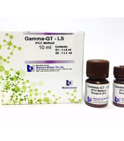
Biotech: GGT / Gamma GT Reagent-biochemistry
Original price was: KSh 5,000.00.KSh 3,600.00Current price is: KSh 3,600.00.Principle of the Test
Most GGT reagents use the Szasz kinetic method, which involves:
-
Substrate:
γ-glutamyl-p-nitroanilide
This compound contains a gamma-glutamyl group. -
Enzymatic Reaction:
GGT in the sample transfers the gamma-glutamyl group to an acceptor (often glycylglycine). -
Product Formation:
The reaction releases p-nitroaniline, which has a strong yellow color. -
Measurement:
The analyzer measures the increase in absorbance at 405 nm.
The rate of color formation is directly proportional to the GGT activity.
Key Reagent Components
The GGT reagent typically contains:
✔ γ-glutamyl-p-nitroanilide
The chromogenic substrate used in the Szasz method.
✔ Glycylglycine (or similar acceptor)
Accepts the gamma-glutamyl group during the reaction.
✔ Buffers
Maintain optimal pH (usually alkaline, around pH 8.2–8.4).
✔ Stabilizers & Preservatives
Ensure reagent stability and long shelf life.
✔ Detergents / Surfactants
Help maintain reagent clarity and enhance reaction performance.


