
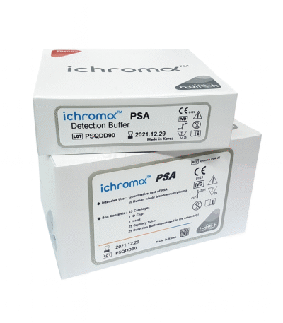
ichroma PSA Prostate-Specific Antigen
ey Features
1. Quantitative PSA Measurement
-
Produces accurate numeric values of PSA concentration.
-
Supports:
-
Prostate cancer screening
-
Monitoring treatment response
-
Post-treatment surveillance
-
Assessment of benign prostatic hyperplasia (BPH) and prostatitis
-
2. Rapid Turnaround Time
-
Results typically available in 10–12 minutes.
-
Ideal for outpatient clinics, urology units, and point-of-care settings.
3. High Sensitivity & Specificity
-
Fluorescence immunoassay technology ensures precise and reproducible results.
-
Good clinical performance comparable to laboratory analyzers.
4. Easy-to-Use Cartridge Format
-
Simple preparation: apply sample → insert into analyzer → get results.
-
Minimal training required.
5. Small Sample Volume
-
Works with whole blood, plasma, or serum.
-
Suitable for fast and convenient testing.
6. Portable & Compact System
-
Compatible with compact ichroma analyzers.
-
Useful in clinics, mobile screening programs, and decentralized testing points.
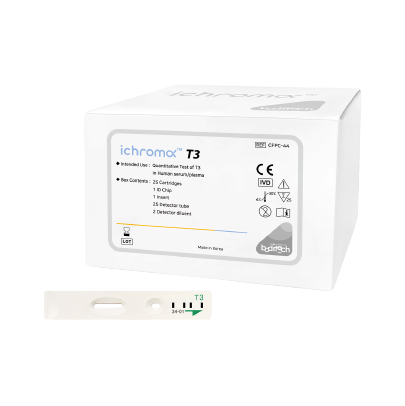
ichroma T3 – Total Triiodothyronine (T3)
Key Features
-
Quantitative T3 measurement: Detects total circulating Triiodothyronine levels for accurate thyroid assessment.
-
Fast turnaround time: Provides results within minutes using automated fluorescence detection.
-
Easy to use: Utilizes a disposable cartridge format compatible with ichroma instruments.
-
Small sample requirement: Typically requires only a small volume of serum or plasma.
-
High sensitivity and specificity: Uses T3-specific antibodies to ensure reliable results.
Clinical Applications
-
Assessment of thyroid disorders, including hyperthyroidism and hypothyroidism
-
Monitoring of thyroid hormone replacement therapy
-
Evaluation of metabolic function related to thyroid hormones
-
Investigation of symptoms such as fatigue, weight changes, and abnormal thyroid-stimulating hormone (TSH) levels
Principle of the Test
The assay uses a fluorescence immunoassay principle:
-
Patient sample is applied to the cartridge.
-
T3 molecules in the sample bind to fluorescently labeled antibodies.
-
The ichroma analyzer measures emitted fluorescence to determine T3 concentration.
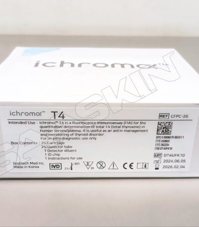
ichroma T4 (Total Thyroxine)
Key Features
1. Quantitative Total T4 Measurement
-
Provides precise numerical values for total T4 concentration.
-
Helps in evaluating:
-
Hypothyroidism
-
Hyperthyroidism
-
Thyroid hormone treatment response
-
Thyroid hormone-binding abnormalities
-
2. Fast Turnaround Time
-
Results available in 10–12 minutes.
-
Ideal for endocrine clinics, general hospitals, and point-of-care testing.
3. Fluorescence Immunoassay Technology
-
Uses fluorescent-labeled antibodies.
-
Ensures high accuracy and strong analytical performance comparable to standard laboratory systems.
4. Easy-to-Use Cartridge Format
-
Simple sample application.
-
Automated reading by the ichroma analyzer.
-
Minimal user training required.
5. Small Sample Requirements
-
Works with whole blood, plasma, or serum.
-
Suitable for routine thyroid evaluation.
6. Compact and Portable System
-
Compatible with lightweight ichroma analyzers.
-
Enables decentralized testing where rapid results are needed.
Clinical Applications
-
Diagnosis of hyperthyroidism and hypothyroidism
-
Monitoring thyroid hormone therapy
-
Evaluation of thyroid gland activity
-
Assessment of thyroid-binding protein disorders
-
Screening in general practice and endocrine clinics
ichroma Testosterone – Total Testosterone Test Cartridge
Key Features
-
Quantitative testosterone measurement: Provides accurate assessment of total circulating testosterone.
-
Rapid testing: Generates results within minutes on ichroma analyzers.
-
Simple workflow: Single-use cartridge designed for easy and reliable operation.
-
High analytical accuracy: Uses testosterone-specific antibodies with fluorescent labeling.
-
Small sample volume: Requires only a small amount of serum or plasma.
Clinical Applications
-
Evaluation of male hypogonadism (low testosterone)
-
Assessment of androgen excess conditions
-
Monitoring testosterone replacement therapy
-
Investigation of infertility in males and females
-
Hormonal profiling in endocrine disorders
-
Supportive test in conditions affecting sexual development and function
Principle of the Test
The assay uses a fluorescence immunoassay method:
-
A serum or plasma sample is applied to the cartridge.
-
Testosterone in the sample binds to fluorescently labeled antibodies.
-
The ichroma analyzer measures fluorescence intensity, which corresponds to testosterone concentration.
Typical Output
-
Quantitative Total Testosterone result displayed on the analyzer
-
Helps clinicians interpret hormonal status along with LH, FSH, SHBG, and estradiol levels
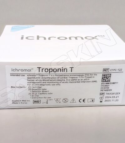
Ichroma Troponin
Key Features
1. Quantitative Troponin I Measurement
-
Provides accurate numerical cTnI values.
-
High clinical relevance for:
-
Early detection of heart attacks (AMI)
-
Risk stratification in chest pain patients
-
Monitoring of myocardial injury
-
2. Rapid Turnaround Time
-
Results are typically available in 10–15 minutes.
-
Ideal for emergency departments, ambulances, clinics, and bedside testing.
3. High Sensitivity & Specificity
-
Fluorescent-labeled antibodies ensure reliable detection.
-
Suitable for detecting both early and late stages of myocardial injury.
4. Easy-to-Use Cartridge Format
-
Simple preparation and loading.
-
Fully automated reading through the ichroma analyzer.
-
Minimal user training required.
5. Small Sample Volume
-
Uses a small amount of whole blood, serum, or plasma.
-
Convenient for emergency or point-of-care settings.
6. Portable and Compact System
-
Works with compact ichroma analyzers for decentralized testing.
Clinical Applications
-
Diagnosis of Acute Myocardial Infarction (AMI)
-
Evaluation of chest pain or suspected cardiac events
-
Monitoring of cardiac injury during treatment
-
Assessment after cardiac surgery or procedures
-
Follow-up of patients with unstable angina
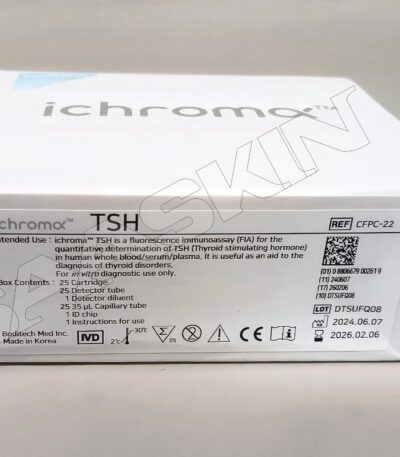
ichroma TSH (Thyroid-Stimulating Hormone)
Key Features
1. Accurate Quantitative TSH Measurement
-
Provides precise TSH values in mIU/L.
-
Supports:
-
Diagnosis of thyroid disorders
-
Monitoring thyroid hormone replacement therapy
-
Screening for congenital hypothyroidism
-
Assessment of pituitary function
-
2. Rapid Turnaround Time
-
Delivers results in 10–15 minutes.
-
Ideal for clinics, endocrine centers, and point-of-care testing.
3. High Sensitivity & Specificity
-
Uses fluorescent-labeled antibodies for strong analytical performance.
-
Comparable to laboratory immunoassay platforms.
4. Easy-to-Use Cartridge System
-
Simple sample loading.
-
Automated reading on the ichroma analyzer.
-
Minimal training required for operation.
5. Small Sample Volume
-
Works with whole blood, plasma, or serum.
-
Suitable for adults, children, and neonatal screening programs.
6. Compact & Portable Testing Platform
-
Compatible with compact ichroma analyzers.
-
Suitable for decentralized healthcare facilities.
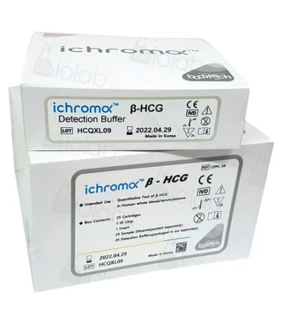
ichroma β-hCG (Beta–Human Chorionic Gonadotropin)
Key Features
1. Quantitative β-hCG Measurement
-
Provides precise numerical values.
-
Clinically useful for:
-
Early pregnancy detection
-
Monitoring pregnancy progression
-
Assessing risk of ectopic pregnancy
-
Monitoring after miscarriage treatment
-
Tumor marker use in certain cancers (e.g., trophoblastic disease)
-
2. Rapid Turnaround Time
-
Results in 10–12 minutes.
-
Suitable for emergency care, maternity clinics, fertility centers, and general practice.
3. High Sensitivity & Specificity
-
Fluorescent-labeled antibody technology ensures accurate detection.
-
Comparable to laboratory-grade immunoassays.
4. Easy-to-Use Cartridge System
-
Simple sample application.
-
Fully automated reading once inserted into the ichroma analyzer.
-
Minimal user training required.
5. Works With Multiple Sample Types
-
Whole blood
-
Serum
-
Plasma
6. Compact & Portable System
-
Ideal for decentralized testing and point-of-care environments.
Clinical Applications
-
Confirmation of pregnancy
-
Monitoring early pregnancy viability and trends
-
Detection and evaluation of ectopic pregnancy
-
Monitoring after medical or surgical management of miscarriage
-
Diagnosis and follow-up of trophoblastic and germ cell tumors
-
Fertility treatment monitoring (IVF/IUI)
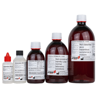
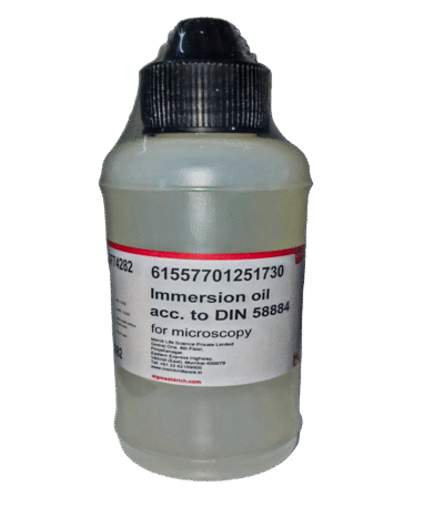
Immersion Oil
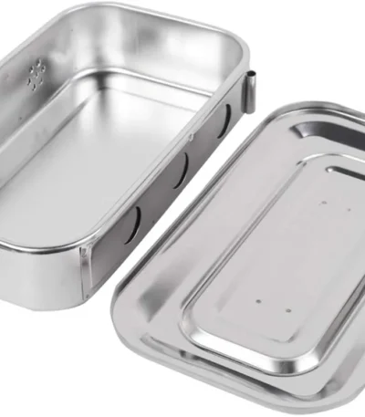
Instrument Tray
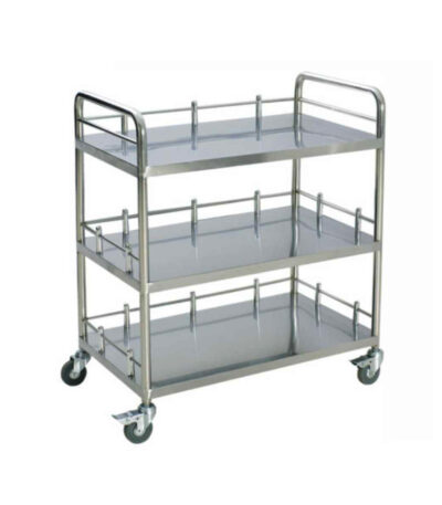

Instrument Trolley
Description:
-
Structure: Usually made of stainless steel or powder-coated metal, providing durability, easy cleaning, and resistance to corrosion.
-
Design: Consists of two or more shelves (tiers) for holding instruments.
-
Mobility: Fitted with four wheels (casters), two of which may have brakes for stability.
-
Handle: Has a side handle for easy movement.
-
Top Shelf: Often used for immediate-use instruments during procedures.
-
Bottom Shelf: Used for storing additional or less frequently used equipment.

Instrument/Medicine trolley
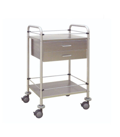
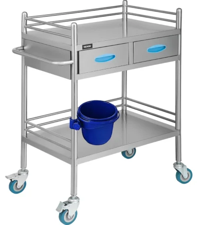
Instrument/Medicine Trolley 2 Shelves/2 Drawers
duct Overview
This mobile instrument/medicine trolley is designed for clinical, hospital, or laboratory settings, offering convenient access, organized storage and smooth mobility. The unit features two open shelves plus two drawers, enabling efficient arrangement of supplies, instruments or medications while maintaining a compact footprint.


